In vivo molecular image evaluation of stem cell migration, distribution and survival in an animal model of epilepsy
RESEARCH PROJECT COMPLETED
Project Coordinator: Jaderson Costa da Costa, MD, PhD < / p>
Members: Mara Lise Zanini, PhD ; Guido Lenz, PhD ; Daniel Marinowic, MSc ; Michelle Domingues, MSc ; Samuel Greggio, PhD ; Gianina Venturin, PhD ; Denis de Assis, PhD and Paola Stradolini , student at the Faculty of Chemistry at PUCRS.
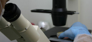
Summary of the Work: the availability of strategies that promote an increase in the number and function of endogenous regenerative cells, or that introduce cells with regenerative potential in the central nervous system (CNS) may represent important alternatives for the treatment of neurological diseases. Thus, stem cell transplantation appears as a promising alternative. However, its clinical application will only be a reality when it is robustly based on experimental data that establish, in addition to effectiveness, the best dose and route of administration, number of applications, as well as the destination and differentiation of transplanted cells. Non-invasive imaging systems, in conjunction with methods for cell labeling, can contribute to the elucidation of these issues. Molecular imaging techniques, such as positron emission tomography for small animals (µPET), allow the presence, distribution, quantity and viability of transplanted cells to be monitored in vivo and in real time (Kircher et al. 2011). In addition, this technology allows sequential studies in the same experimental model, facilitating longitudinal designs.
Monitoring the distribution and migration of biologically active cells in a living organism is crucial for the development of cellular therapies. Thus, the use of µPET represents an important research tool. The study proposes the application of this technology to monitor in vivo the viability, migration and survival of stem cells transfected with an HSV1- sr39tk gene and transplanted into an animal model of epilepsy, using the µPET / CT technique and use of the radioisotope [18F] -FHBG (9- (4- [18F] Fluorine-3-hydroxymethylbutyl) guanine). In order for these cells to be detected, they will be marked indirectly by the introduction of an enzyme-based reporter gene, in which the enzyme product will phosphorylate an exogenous substrate marked with Fluorine-18.
Research areas involved: CPR, CPPC, Neuroscience and Cell Signaling Laboratories - IPB, Laboratory of Cell Signaling and Plasticity - UFGRS.
Funding entity: CNPq - Edital Universal 2012